HOMEOSTASIS
How Nerve Signals Maintain Homeostasis
nervous_system3.jpg

Divisions of the Nervous System

The nervous system has two main divisions: the central nervous system and the peripheral nervous system. The central nervous system consists of the brain and the spinal cord, and the peripheral nervous system consists of nerves and ganglia.  The peripheral nervous system is subdivided into somatic and autonomic nerves. The somatic nerves control the skeletal muscles and the autonomic nerves control internal organs. The autonomic system has two parts; the sympathetic, which deals with emergency situations and the parasympathetic for everyday activities. 


Anatomy of a Nerve Cell

In the nervous system there are two different types of cells: glial cells and neurons. Glial cells are nonconducting cells and are important for the structural support and metabolism of the nerve cells. Neurons are the functional units of the nervous system. 


Neuron Anatomy and Function 

Neurons are nerve cells that conduct nerve impulses. There are three types of neurons:

  • sensory neurons (afferent - conducting impulses towards the central nervous system)
  • interneuron (association neurons)
  • motor neuron (efferent - conducting impulses from the central nervous system)

Every neuron consists of three components

  • cell body - carries out all cell functions
  • dendrite - conducts nerve impulses towards the cell body
  • axon - conducts nerve impulses away from cell body

Many axons are covered with a white coat of fatty protein called the myelin sheath.  The type of glial cell that produces the myelin sheath is called Schwann cells.  The areas between sections of myelin sheath along the axon are known as nodes of Ranvier.  Nerve impulses jump from one node to another, which speeds up the movement of nerve impulses.


neuron1.jpg


Simple Reflex: Reflex Arc

Reflex arcs involve five essential components: the receptor, the sensory neuron, the interneuron in the spinal cord, the motor neuron, and the effector (muscles).  Most reflexes occur without brain coordination. 

For example, if you burn your hand on a hot iron, the sensation of heat is detected by receptors in your skin, and an impulse is initiated in a sensory neuron.  The sensory neuron carries the impulse to the spinal cord and passes it to a interneuron. Then, the interneuron transmits the impulse to the motor neuron.  The motor neuron causes the muscles in your hand to contract and pull away.  This process occurs in less than a second, before even the brain is informed.  Reflexes are involuntary and often unconscious.

The brain becomes aware of an automatic refelx action when the sensory neurons pass impulses to various interneurons, some of which send impulses to the brain.


Nerve Impulse Transmission

A nerve impulse is an electrochemical event involving the movement of unequally distributed ions across the nerve cell membrane.

The following table explains what occurs in three different stages in a neuron:


A Neuron at Rest:


·       Sodium ions (Na+) remain in a greater concentration outside the neuron 

·       Potassium ions (K+) are in a greater concentration inside the neuron

·      The cell membrane is impermeable to Na+

·       The unequal distribution is maintained by a sodium-potassium pump

·       The membrane potential inside the nerve cell is negative (approx. -60 millivolts), caused by the large negatively charged proteins

·       This is called resting potential


A Stimulated Neuron:


·       The stimuli could be electrical, mechanical, or chemical

·       Membrane becomes permeable to Na+

·       The Na+ rush into the nerve cell through protein channels, known as ion gates

·       The membrane potential inside the cell becomes positive (approx. +40mv) relative to the outside of the cell

·       This is called depolarization, or charge reversal, and it is the nerve impulse


Restoring a Neuron:


·       As Na+ diffuses into the cell, K+ begins to diffuse out of the cell, restoring the membrane potential to -60mv

·       This is called repolarization

·       The sodium-potassium pump moves Na+ out of the cell and K+ into the cell through the same type of protein channels

·       ATP is used as the energy to maintain the pump

·       When the sodium-potassium distribution has returned to normal, the membrane is said to have recovered


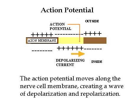
Movement of the Action Potential

The movement of sodium ions into the nerve cell causes a depolarization of the membrane and this signals an action potential. The areas adjacent to the depolarized membrane are in turn affected, resulting in the movement of the impulse along the axon.


actionpotential2.jpg

Synaptic Transmission


Small spaces between neurons are known as synapses.  Small vesicles containing chemicals called neurotransmitters are located at the end plates of axons.  When a nerve impulse reaches the end of an axon, the neurotransmitters are released from the presynaptic neuron (the axon terminal) and diffuse across the synaptic cleft and attach themselves to receptor sites on the membrane of the postsynaptic neuron (the dendrite).  The action potential continues along the postsynaptic neuron. 


Acetylcholine is an example of a neurotransmitter found in the end plates of many nerve cells.

synaptic2.gif

The Central Nervous System

The Spinal Cord

The spinal cord carries sensory nerve messages from receptors to the brain.  The brain passes the message to motor nerves, which activate effectors - muscles, organs, and glands.

The Brain

Brain

Function

Cerebrum

·        Stores sensory information and initiates voluntary motor activities

·        The largest and most highly developed part of the brain


Cerebral cortex

·        The outer lining of the cerebral hemispheres


Corpus Callosum

·        A nerve tract that joins the left and right cerebral hemispheres


Thalamus

·        Area of the brain that coordinates and interprets sensory information and directs it to the cerebrum


Cerebellum

·        The region of the brain that coordinates muscle movement


Medulla Oblongata

·        The site of autonomic nerve control (internal organs)


Pons

·        The region of the brain that acts as a relay station by sending nerve messages between the cerebellum and the medulla oblongata


Hypothalamus

·        Responsible for coordinating many nerve and hormone functions

·        Controls homeostasis


Pituitary

·        Functions as a control centre, coordinating the endocrine and nervous system

brain1.gif

Home

Maintaining an Internal Balance | The Importance of Excreting Wastes | Chemical Signals Maintain Homeostasis | How Nerve Signals Maintain Homeostasis | The Maintenance of Balance by the Immune System | Key Terms | Quiz | Test

Enter supporting content here